
HIGH-TECH TIME TRAVEL
Accurately dating and identifying a painting that is nearly 400 years old is difficult, but thanks to the efforts of Utah State University staff, an art conservator and the high-tech support of Logan Regional Hospital, that’s exactly what was attempted July 13.
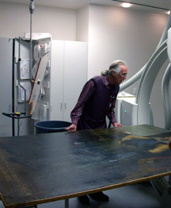
The patient, a nearly five-foot by seven-foot plus painting of undetermined European background, was transported to Logan Regional Hospital a service of Intermountain Health Care, by the university’s moving crew headed by Chet Smith. The patient was a little soiled and suffered a few cracks, but it was the bones of the work that interested Rose Milovich, Nelson Ahrnsbrak and Steven Prins. Milovich is the art and book art curator for University Libraries in Special Collections and Archives, Ahrnsbrak is her assistant and Prins is an independent art conservator from Santa Fe, N.M. The team hoped to pick up clues at the hospital to accurately date the painting and, perhaps, identify the artist who created the work in the 17th century.
Through the emergency room doors the painting traveled, down the hallway, finally making its way to the Special Procedures Lab, which normally sees slightly younger cardiac patients. Into the cath lab and onto the table the artwork went, ready for digital imaging under the supervision of Rod Cevering, a special procedures radiology technician at Logan Regional. All was ready for the high-tech adventure that would, with hope, take the Utah State team time-traveling, solving a few mysteries along the way.
Supporting Milovich in the project were others at the hospital, including Danette Steinitz and cath lab manager Carol Chambers.
Once on the table, the painting was carefully manipulated by Prins, positioning the work so hidden details could be seen. Using a penny as a marker to note grids, the painting was carefully scanned section by section. Images were recorded at the lowest kilovoltage possible (in this case, 50 kv), to show the subtle differences in densities of the work. Images began to pop up on the monitor. There were the nails used to attach the canvas to the stretchers or frame. Canvas differences could be seen. Spots, blotches and imperfections hidden for hundreds of years flashed on the screen.
X-ray technology is used much the same in the art world as it is with the body. It can penetrate to reveal hidden elements of structure and detail. It can detect previous alterations and repairs. X-ray is one piece of the puzzle in identifying and dating art works. It can help “see” the structure, condition and attributable patterns of execution by the artist.
After a preliminary review of the images shot at Logan Regional Hospital, Prins said he was pleased. His review of the images reinforces many suspicions from the visual inspection
The artwork was originally donated to the university by L. Boyd and Anne McQuarrie Hatch and has been housed in the Merrill Library’s Hatch Room. Conservator Prins has been brought in to evaluate the work, along with a smaller landscape by Jules Dupre (French Barbizon School), and begin restoration work. The process is in anticipation of the move to the university’s new library, currently under construction.